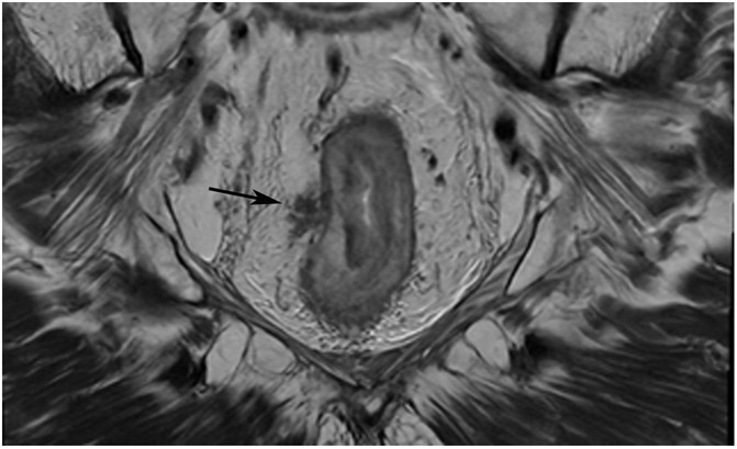
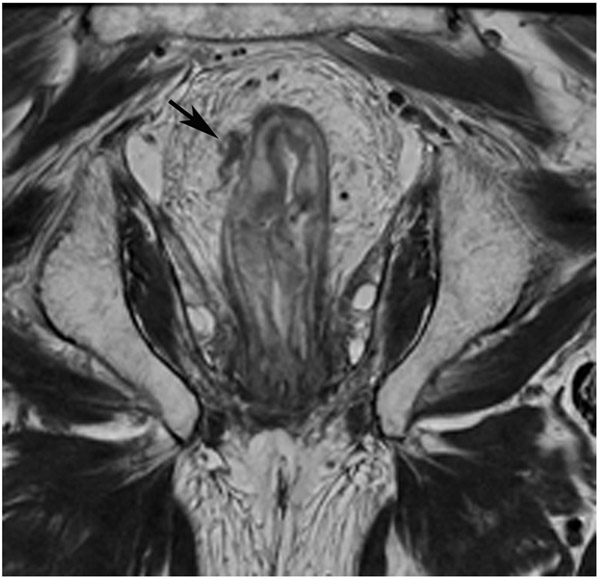
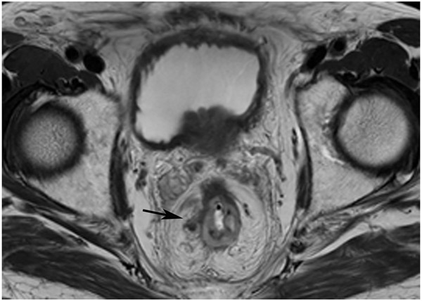
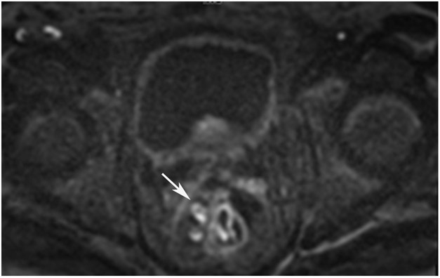
Figure 25:
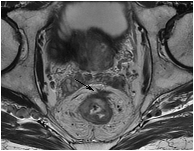
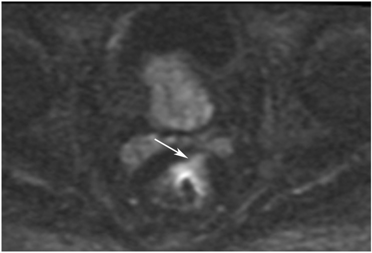
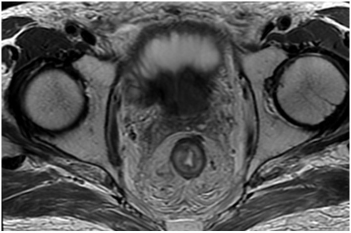
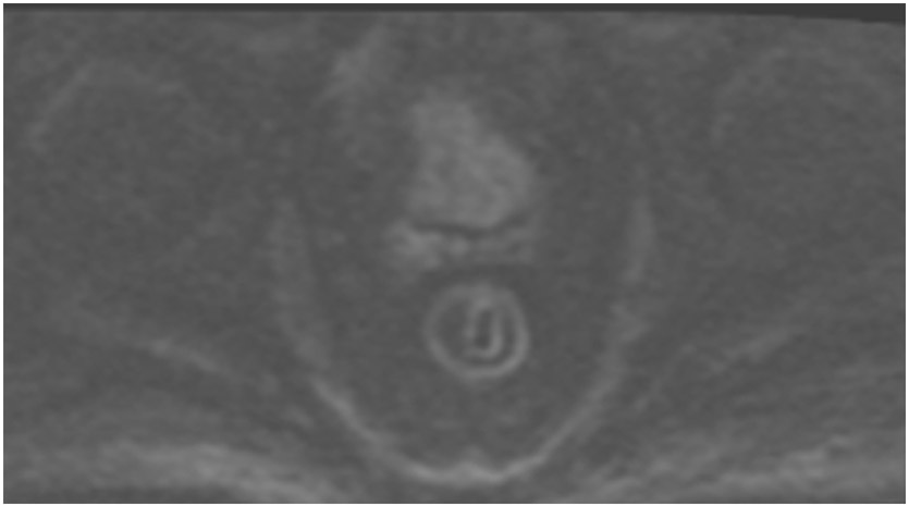
86-year-old man with primary rectal cancer 5.9 cm from the anal verge. (a) Axial T2WI reveals a tumor extending to the mesorectal fascia (”circumferential resection margin”) anteriorly at 12–1 pm (arrow). (b) Axial b1500 FOCUS reveals diffusion restriction in the intramural and extramural tumor (arrow). (c) 8 months from baseline, post-TNT axial T2WI reveals minimal scarring in the tumor bed and in the mesorectal fat. (d) b1500 FOCUS DWI shows no restriction. (e–h) Surveillance MRI 1 year later reveals irregular node/tumor deposit or extramural venous invasion (EMVI) (e; arrow, oblique axial, f; arrow, oblique coronal, g; arrow, straight axial). There is also DWI restriction (h; arrow), suspicious for EMVI. The patient subsequently developed liver metastases (very common in EMVI cases).
TEACHING POINT: Tumor regrowth may occur outside the primary tumor bed in up to 5% of cases as either lymph node invasion, EMVI, or tumor deposit. Remember to look in the mesorectal fat.

