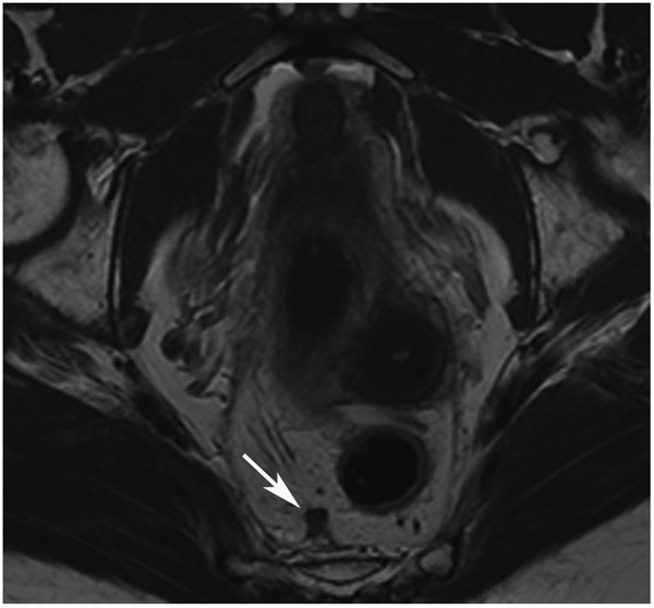
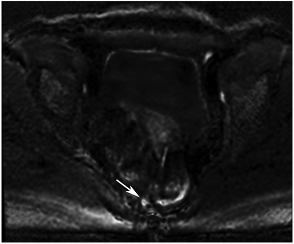
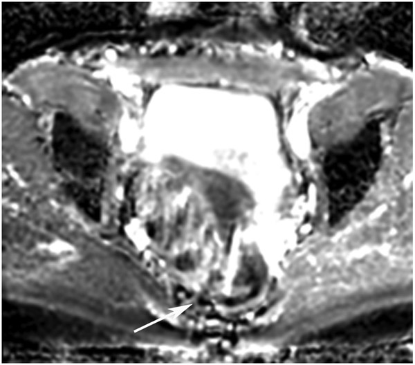
Figure 26:
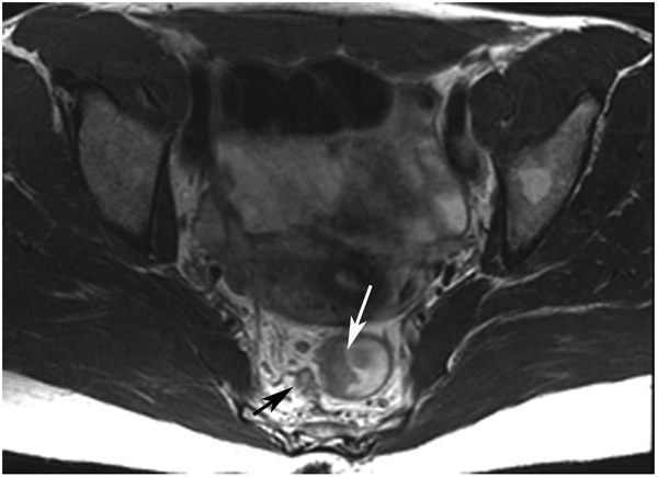
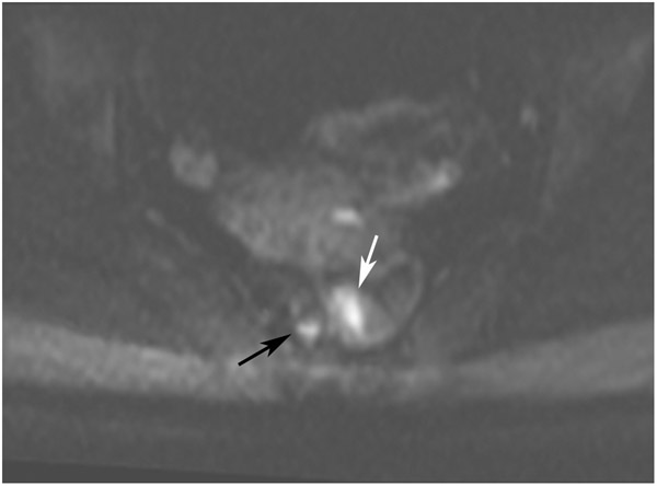
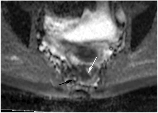
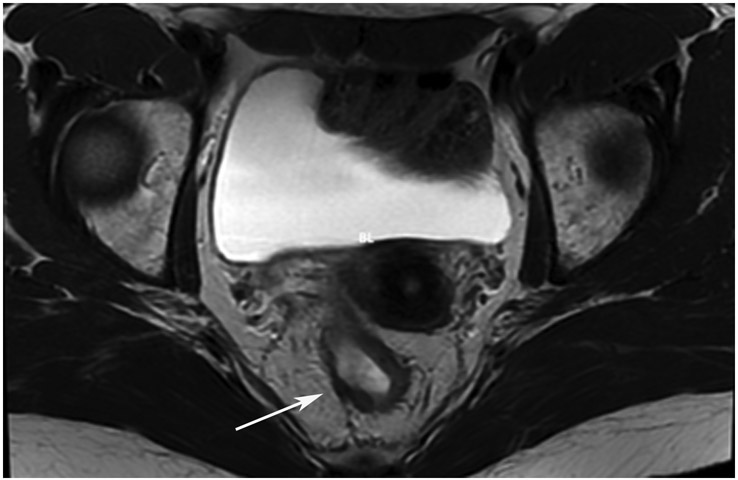
45-year-old woman with primary rectal cancer 10 cm from the anal verge. (a–c) Axial T2WI MRI (a), b800 DWI (b), and ADC (c) reveal a partly circumferential mass (long arrows) and discontiguous extramural venous invasion/tumor deposit (shorter arrows) showing expected diffusion restriction in the true tumor. (d–e) 15 months later, following induction chemotherapy and short-course radiation (TNT), the tumor disappeared while a scar appeared (arrow, d) and there was no restriction on DWI (e). (f–h) Oblique axial T2WI as well as b800 DWI and ADC images reveal regrowth in a discontiguous tumor nodule or focus of EMVI (arrows).
TEACHING POINT: Tumor regrowth may occur outside the primary tumor bed in up to 5% of cases as either lymph node invasion, EMVI, or tumor deposit. Remember to look in the mesorectal fat.

