Figure 27:
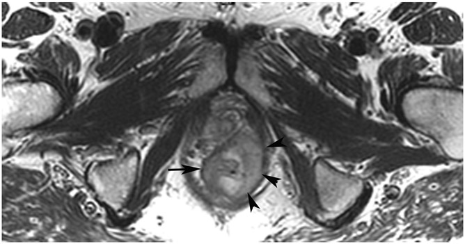
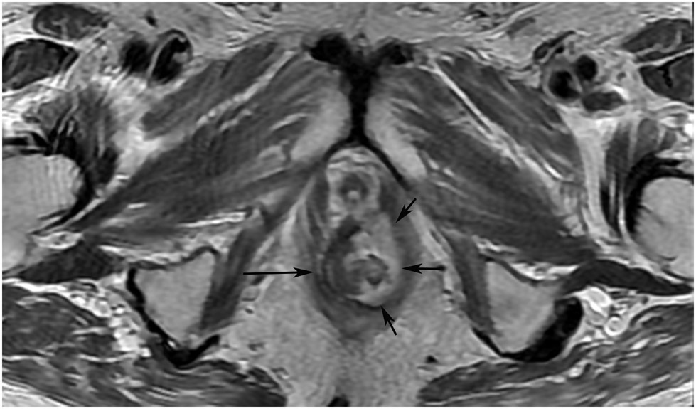
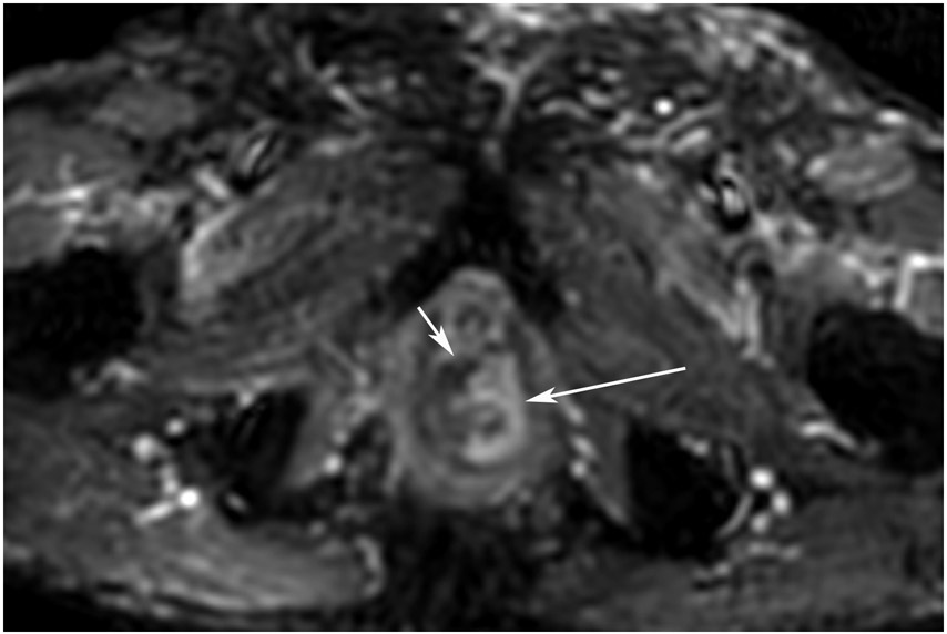
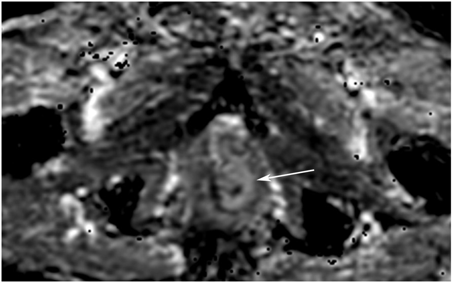
A 68-year-old man with Crohn’s Disease with chronic fistulae developed mucinous cancer which was treated with TNT. (a) Baseline MRI shows a tumor with intermediate signal (arrow), with higher signal in the mucinous component (arrowheads). (b) 8 months post-TNT axial T2WI shows a dark scar at 7–11 pm (long arrow) and mucinous degeneration (small arrows). (c) Axial b800 DWI reveals a dark signal in the scar (short arrow) and a bright signal (long arrow) which is T2 shine-through as confirmed on the ADC map. (d) ADC map shows a bright signal in the mucinous area due to T2 effects (arrow). The patient required pelvic exenteration. Pathology showed 99% treatment response. He has no evidence of disease 7 years later.
TEACHING POINT: Mucin may be cellular (tumor present) or only acellular. Because of the high water content of mucin, the T2 effect may overshadow any restriction and miss viable tumor. In such cases, a disclaimer is recommended to say that “MRI cannot distinguish cellular from acellular mucin.” Of course, if there is another restricting component in the tumor, the point is moot.


