Figure 28:
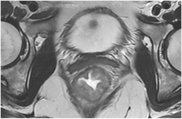
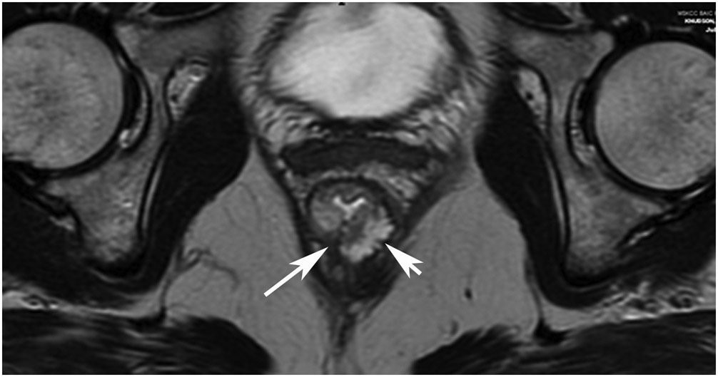
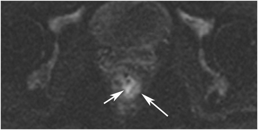
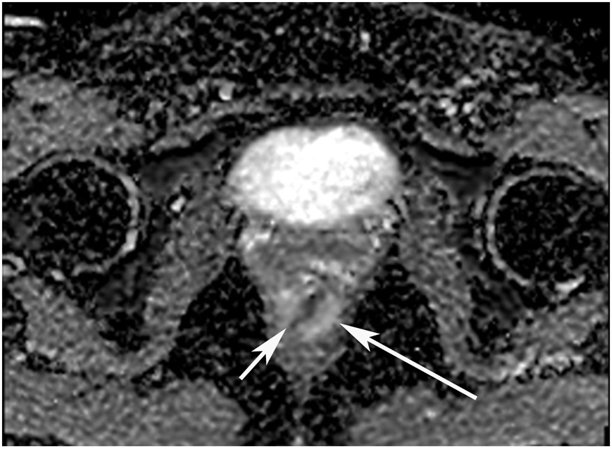
45-year-old woman with a dMMR (deficient mismatch repair) circumferential tumor 2.5 cm from the anal verge who participated in an immunotherapy trial (Dostarlimab). (a) Axial T2WI shows a low circumferential intermediate-T2-signal tumor. (b) 3-month axial T2WI reveals mucinous degeneration of much of the tumor deep in the wall (short arrow) and some residual intermediate wall/tumor more superficially (long arrow). (c) On b1500 DWI, there is very high signal adjacent to but not deriving from the collapsed lumen (short arrow) and less bright signal deeper in the wall (long arrow). (d) On the ADC map, the inner dark signal (short arrow) indicates tumor corresponding to the inner very bright DWI signal, while the outer bright signal indicates T2 shine-through from mucin (long arrow). The patient continues on routine follow-up per trial requirements. PET scans were also done, indicating partial response (not shown). A recent series of similar patients published in NEJM has shown 100% response rates [70].
TEACHING POINT: Although many cases of mucin after treatment only show T2 shine-through (see prior case in Figure 27), there may still be residual tumor that we cannot detect. Some cases upon detailed close inspection will still have DWI restriction and it would be an oversight not to carry through with standard analysis with DWI/ADC.


