FIGURE 4:
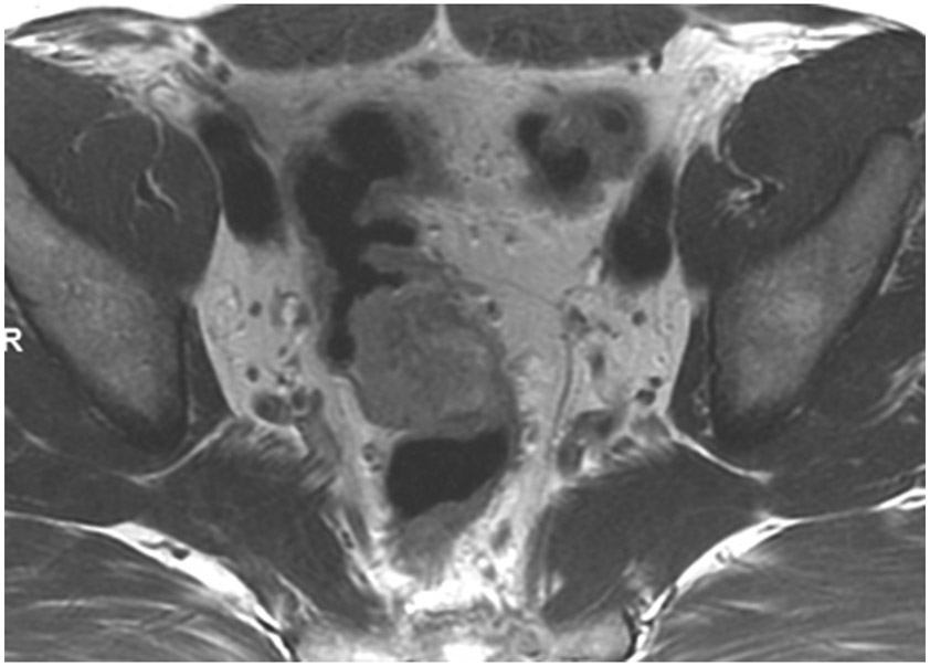
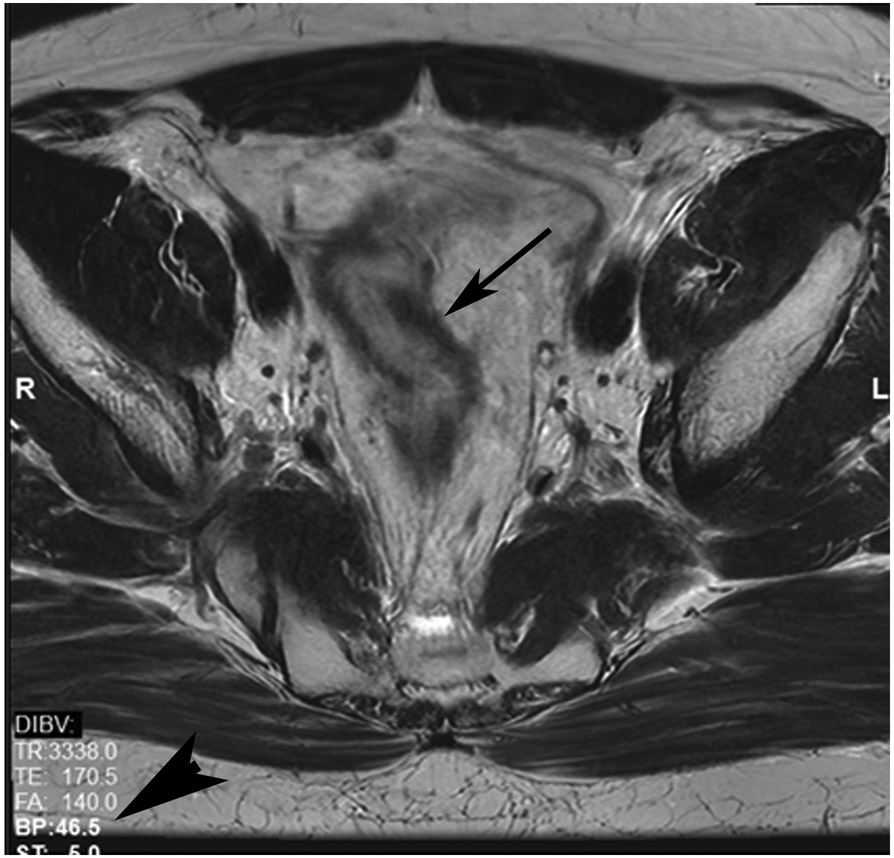
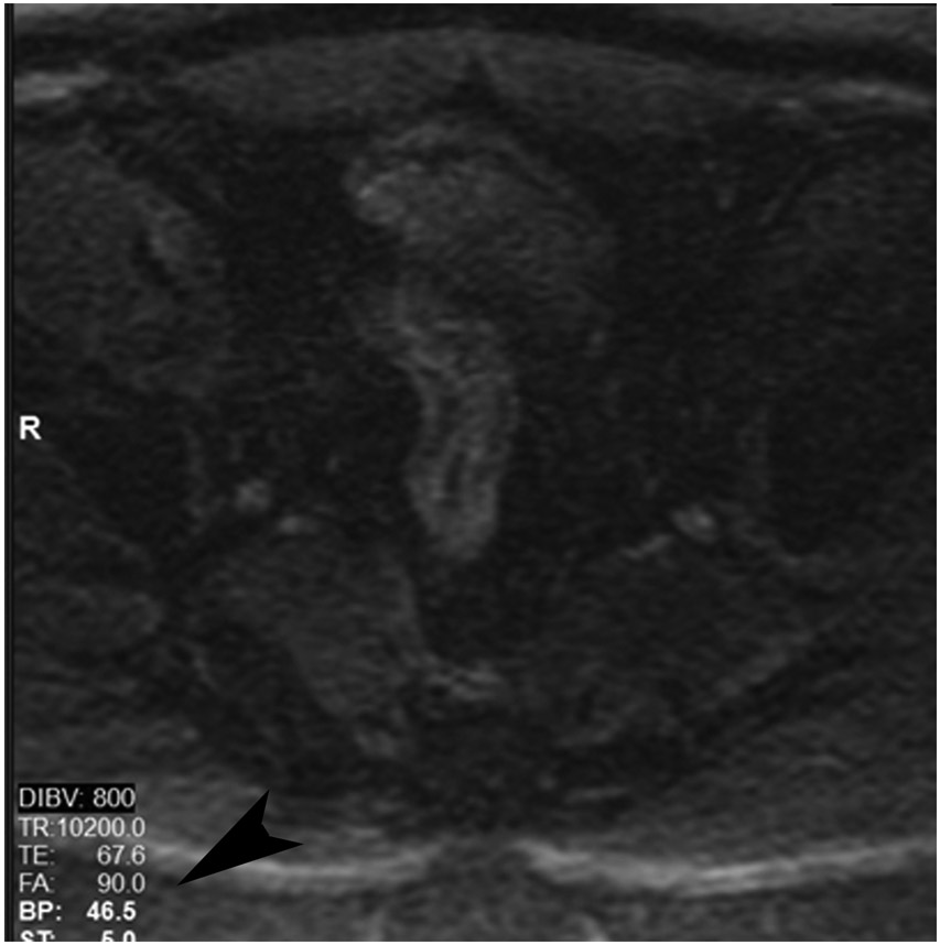
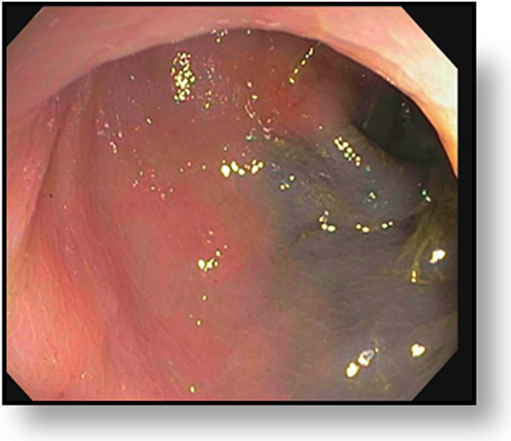
cCR at the time of first post TNT MRI in a 52-year-old man with T3N+ rectal cancer 14 cm from the anal verge. (a) 5-mm straight axial MRI slice through the tumor bed reveals a T2-intermediate-signal tumor. (b) 5-mm straight axial MRI slice at 9 months (2 months post end of TNT) shows the disappearance of the tumor and the appearance of a dark signal intensity scar at location of tumor. This attachment point is a little thicker than elsewhere (arrow). Note the bed position of 46.5 (arrowhead). (c) Matching 5-mm straight axial b800 MUSE [multiplexed sensitivity encoded] DWI slice reveals some striated signal of normal wall but no extra signal at tumor site. Note the bed position of 46.5 (arrowhead). (d) Endoscopy shows no tumor (blue tattoo material incidentally noted).
TEACHING POINT: A scar will be located where the tumor was attached to wall. On DWI images (b800 and/or b1500), the same bed position should show no extra signal compared with the background wall. When this is true, the MRI report can state “clinical complete response.”


