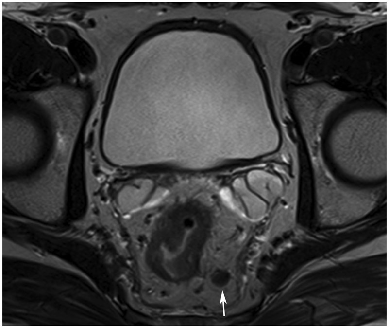
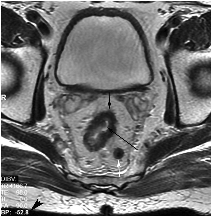
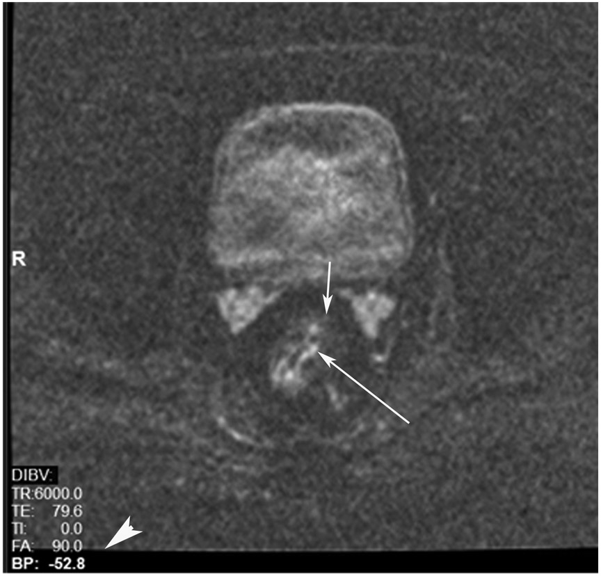
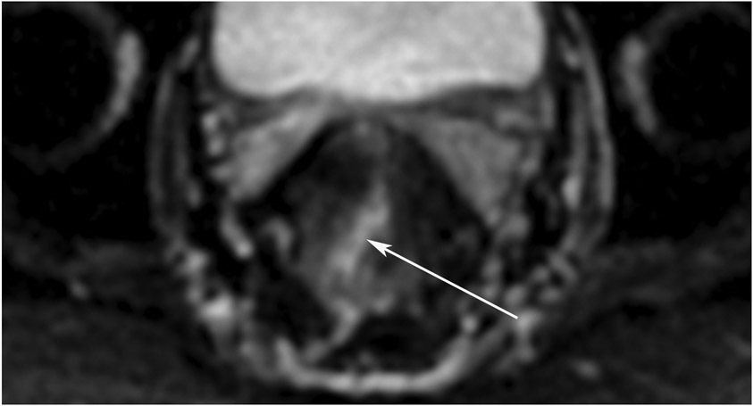
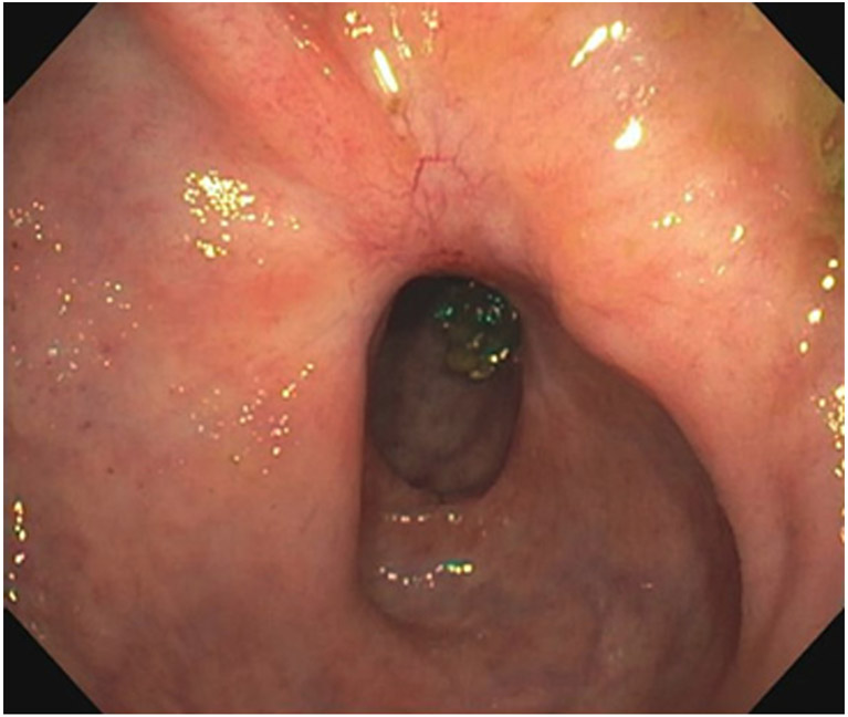
FIGURE 5:
cCR at the time of post-chemoradiotherapy (CRT)-only MRI and before consolidation in a 48-year-old man with T3N+ rectal cancer 5 cm from the anal verge. (a) 5-mm straight axial MRI slice through the tumor bed reveals a circumferential tumor with intermediate T2 signal. (b) 5-mm straight axial MRI slice at 3 months (3 weeks post end of CRT) shows the disappearance of the tumor and the appearance of a dark signal intensity scar at the location of the tumor (arrow). Note the bed position of −52.8 (arrowhead). Fluid in the lumen is present (long arrow). (c) Matching 5-mm straight axial b800 DWI slice reveals no DWI signal in the wall. There is expected signal in the lumen as was seen on T2WI (long arrow). Note the bed position of −52.8 (arrow). (d) ADC map shows a bright signal in the lumen, proving fluid in the lumen rather than tumor with restriction. This is T2 shine-through (long arrow). (e) Endoscopy shows no tumor. The patient still had clinical complete response at 14 months after the first MRI.
TEACHING POINT: ADC maps must always be used when there is a question of restriction to ensure that it is not due to a T2 effect. Finally, this case is unusual in that the nodes remained over 0.5 cm (white arrows in a and b), not considered sterilized by MRI, but the patient continued undergoing the Wait-and-Watch approach for another year without evidence of disease.

