FIGURE 7:
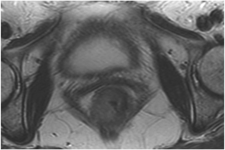
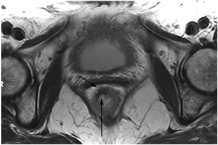
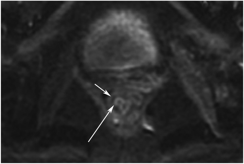
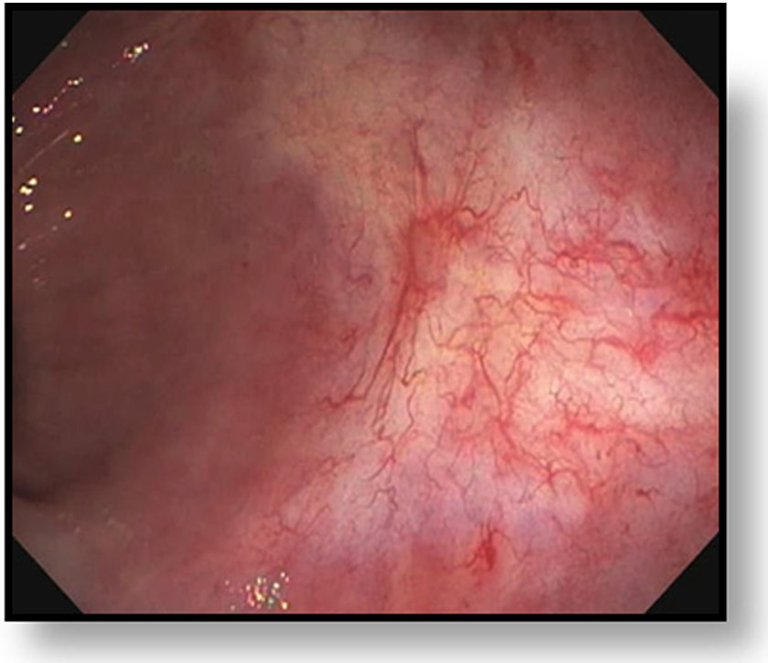
cCR 5 years into surveillance in a 71-year-old woman with T3Nx rectal cancer 4 cm from the anal verge. (a) Straight axial 5-mm slice MRI through the tumor bed reveals a partly circumferential tumor with intermediate T2 signal. (b) 5-mm straight axial MRI slice at 5 years. Note the dark signal intensity scar at the location of the tumor (arrow). Note that next to the scar, the lumen is a bit ballooned out from atrophy or healed ulceration (long arrow). This is common, but the overlying wall has no DWI signal (see part c). (c) Matching 5-mm straight axial b1500 FOCUS DWI slice reveals a scar, with no restriction in the scar itself (arrow). Extra internal signal is from mucosa (long arrow). Always confirm T2 shine-through with the ADC map. (d) Endoscopy shows a flat whitish scar with telangiectasias, which is one appearance of cCR along a spectrum. The patient remains free of disease at 6 years.
TEACHING POINT: A scar will be located where the tumor was attached to wall. On DWI images (b800 and/or b1500), the same bed position should show no extra signal compared with the background wall. When this is true, the MRI can state “clinical complete response.” ADC maps must always be used when there is a question of restriction to ensure that it is not due to a T2 effect.


