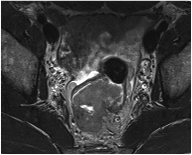
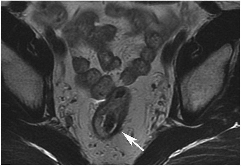
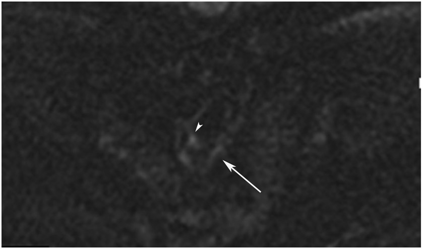
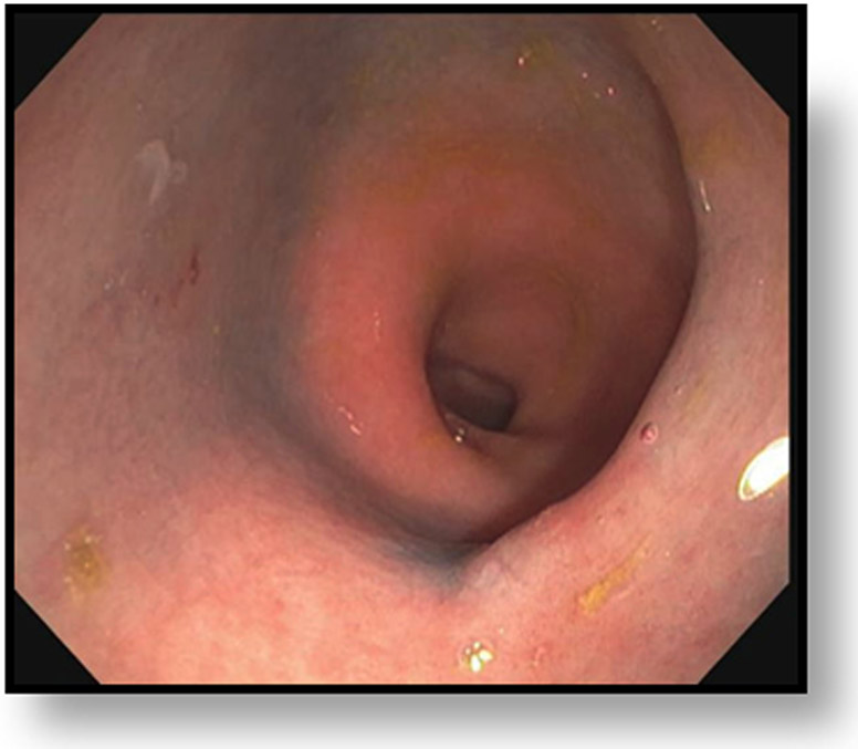
FIGURE 8:
cCR 1.5 years into surveillance in a 45-year-old woman with T3N+ rectal cancer 8 cm from the anal verge. (a) 5-mm straight axial MRI slice through the tumor bed reveals a T2-intermediate-signal bulky circumferential tumor. (b) 5-mm straight axial MRI slice at 1.5 years shows a scar at the location of the tumor (arrow). Note the edema on the opposite wall from radiation to normal mucosa. (c) Matching 5-mm straight axial b1500 FOCUS DWI slice shows no obvious additional signal compared with that of the normal wall (arrow) (please note the focus of signal on the opposite wall [white arrowhead] that is likely T2-shine through from edema as seen in (b)). (d) Endoscopy shows no tumor regrowth.
TEACHING POINT: Note that the normal rectal wall can show variable amounts of DWI signal, but it is extra signal which would raise suspicion. Of course, this subjective determination is the challenge in these cases.


