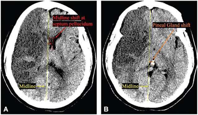
Fig. 1.
Midline shift at septum pellucidum and pineal gland shift on non-contrast CT scans. (A) Midline shift (red line) at the level of septum pellucidum (red dotted line). (B) Pineal gland shift (orange line) at the level of pineal gland (orange dotted line) have been associated with worsened outcome and arousal in large hemispheric infarction.

