Figure 3.
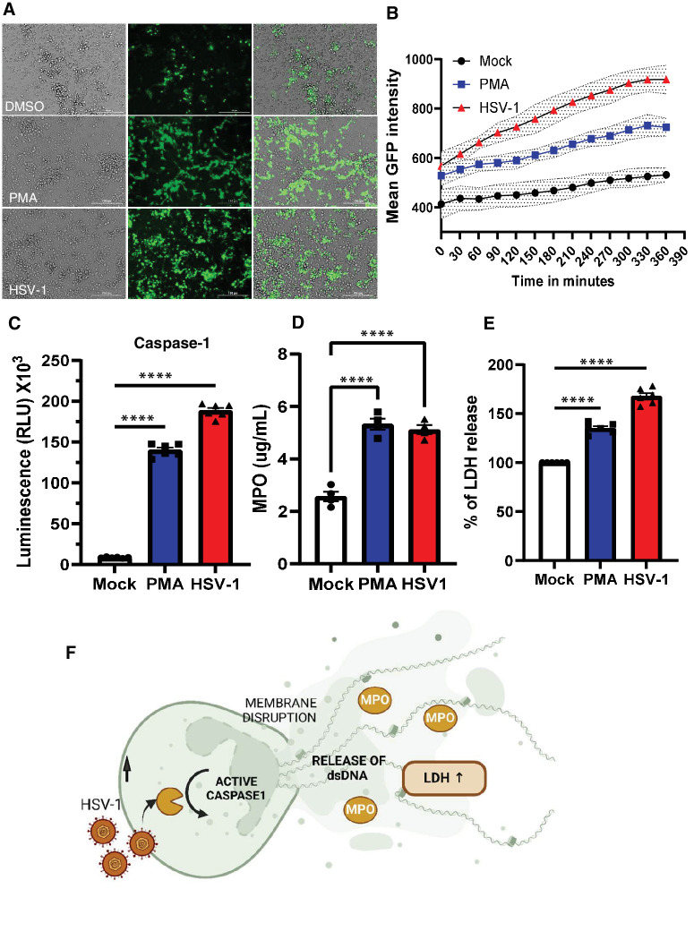
HSV-1 infection triggers rapid NETosis in neutrophils. (A) Fluorescence imaging of mock, PMA treated, and HSV-1 infected neutrophils. Cells were treated with Sytox green reagent (Scale bar = 100 µm). (B) Quantification of mean green fluorescence intensity over time for live cell imaging of mock, PMA treated, and HSV-1 infected neutrophils. Each condition (3-4 replicates per group) with error bands. Time interval was 30 minutes. (C) Specific and rapid detection of caspase-1 activity was measured by bioluminescent assay. Caspase-1 activity was measured in cell supernatants collected from different conditions. (D) Quantitative MPO assay for mock, PMA treated, and HSV-1 infected neutrophils after 1 hour (n = 4 each group). (E) LDH release in the supernatant was measured from the mock, PMA treated and HSV-1 infected samples. (F) Overall schematic of cell death trigger and release of NETs. Activation of caspase-1 followed by release of cellular content and dsDNA in the surrounding medium. One-way ordinary ANOVA was performed for statistical analysis. ****P < 0.000.

