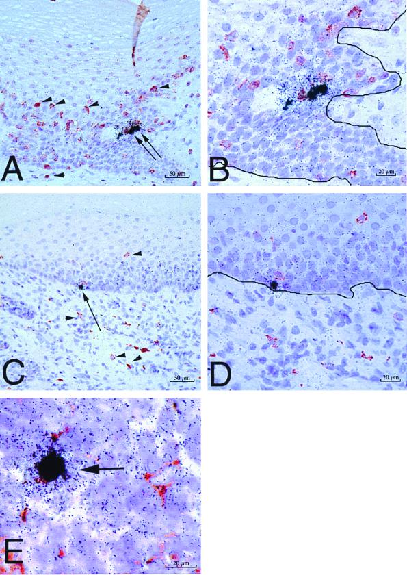
FIG. 3.
SIV-infected cells in tissues of rhesus macaques as detected by combined ISH and immunohistochemistry. SIV RNA+ cells in formalin-fixed sections of vagina at 24 h PI are shown. (A) SIV RNA+ cells (arrows) in the vaginal epithelium and uninfected CD3+ T cells (red, some are denoted by arrowheads) in the vaginal epithelium and lamina propria (animal 24294, 24 h PI). (B) Higher-magnification view of panel A. Note that the SIV RNA+ cells are not CD3+ T cells. The solid black line demarcates the basal lamina. (C) SIV RNA+ cell (arrow) in the basal layer of the vaginal epithelium and uninfected HAM 56+ macrophages (red, some are denoted by arrowheads) in the vaginal epithelium and lamina propria (animal 24294, 24 h PI). (D) Higher-magnification view of panel C. Note that the SIV RNA+ cell is not a macrophage and the macrophages are not SIV RNA+ cells. The solid black line demarcates the basal lamina. (E) SIV RNA+ macrophage (arrow) in the paracortex of an iliac lymph node (animal 24294, 24 h PI). Note in panels A and C that the vaginal epithelium is intact at 24 h PI, consistent with the atraumatic virus inoculation procedure. ISH using 35S-labeled SIV riboprobes combined with immunohistochemistry using AEC as the chromogen and Meyer's hematoxylin counterstain were used.

