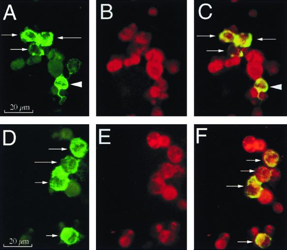
FIG. 4.
Immunophenotypic characterization of SIV RNA+ cells in HLA-ECS cytospin slides of vaginal epithelium at 18 h PI. Panels A to C show A single, high-magnification field in a cytospin slide from animal 23319 (18 h PI). (A) Viewed through an appropriate band-pass filter, SIV RNA+ cells were detected (green, arrows), and some of these cells had dendritic processes (arrowhead). (B) Most cells express p55+ (red), a marker for DC. (C) Viewed through a double band-pass filter, all the SIV RNA+ cells in this field are p55+ DC (arrows). Panels D to F show a single, high-magnification field in a cytospin slide of vaginal epithelium from animal 23319 (18 h PI). (D) SIV RNA+ cells (green, arrows). (E) Most cells (red) express CD1a, a marker for LC. (F) Viewed through a double band-pass filter, all the SIV RNA+ cells in this field are CD1a+ LC (arrows). Combined ISH (digoxigenin-labeled riboprobe, Tyramide-FITC detection system) and immunofluorescent antibody labeling of cell markers (Texas red detection system) were used.

