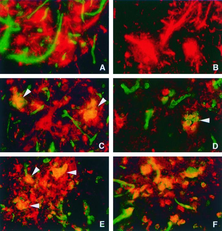
FIG. 1.
Double immunofluorescence microscopy in the CNS of C57BL/6 mice (A, C, D, E, and F) and B2m-KO mice (B) after infection with NSV. Panels A, B, C, and D show double staining for MHC class I (H-2Kb/H-2Db) antigen (green) and SV antigen (red). Panel E shows double staining for F4/80 antigen (green) and SV antigen (red). Panel F shows double staining for H-2Kb/H-2Db antigen (green) and F4/80 antigen (red). At 3 days after infection, H-2Kb/H-2Db antigen was detected in endothelial cells of C57BL/6 mice (A) but not in B2m-KO mice (B). SV-antigen-positive neurons shown in panel A (red) did not demonstrate MHC class I immunoreactivity. At 5 days after infection (C and D), the numbers of MHC class I immunoreactive cells increased, and cells that were positive for MHC class I and SV (arrowheads) were detected. Note that SV-antigen-positive cells with neuronal morphology do not show immunoreactivity for MHC class I. At 5 days after infection (E), F4/80-antigen-positive cells (green) with engulfed SV antigen (red) accumulated. Most F4/80-positive cells (red) detected in NSV-infected foci show MHC class I (green) immunoreactivity (F). (A, B, C, E, and F) Ventral horn of lumbar spinal cords; (D) thalamus. (A, B, C, D, and F) Magnification, ×322; (E) magnification, ×403.

