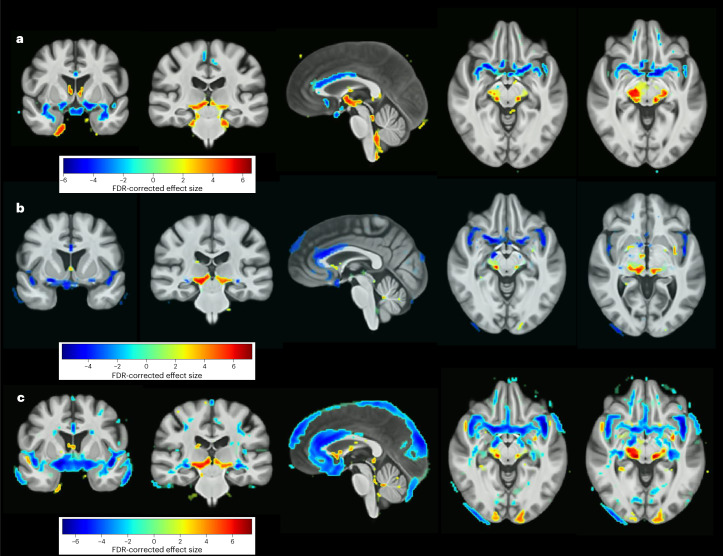
Fig. 5. Neuroanatomical case-control differences.
a,b, FDR-corrected voxel-wise comparison of gray-matter volume differences between the whole MDD participant group versus healthy controls (a) and after controlling for medication status (b). c, Gray-matter volume differences between MDD participants in a first episode of depression and healthy controls. The color bars indicate the strength of the group differences (MIDAS statistic) between MDD and healthy control participants.

