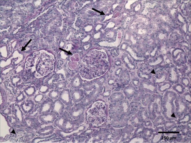
FIGURE 1.

Light microscopy of kidney biopsy. Cortical zone showing three glomeruli with normal architecture, mild hypertrophy, and no evidence of proliferative, inflammatory, necrotising, or sclerotic lesions. The tubules reveal irregularities along the luminal epithelial border, severe flattening, distension, and intraluminal debris, consistent with signs of acute injury (arrows). There are scattered lymphocytic cells at interstitium (arrow heads). Brown tubular casts are not recognized (Periodic acid‐Schiff stain, original magnification ×100).
