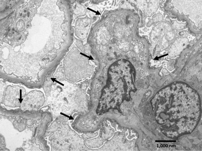
FIGURE 2.

Electron microscopy of kidney biopsy. Segment of a glomerulus with four capillary loops showing podocytes with complete foot processes effacement, swollen cytoplasm and condensed cytoplasmic filaments over the underlying basement membrane (arrows), consistent with a minimal change type lesion. Electron‐dense deposits are not observed, and the glomerular basement membranes appear normal (uranyl acetate‐lead citrate, original magnification ×7,200).
