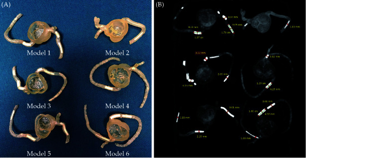
Figure 25.
Three-dimensional printed patient-specific coronary models based on the simulation of calcified plaques in the coronary arteries.
(A): Three-dimensional printed models (n = 6) with simulated calcified plaques in coronary artery branches; (B): measurements of plaque dimensions on 2D maximum-intensity projection images using 0.5 mm slice thickness. Reprinted with permission under open access from Sun, et al.[153]

