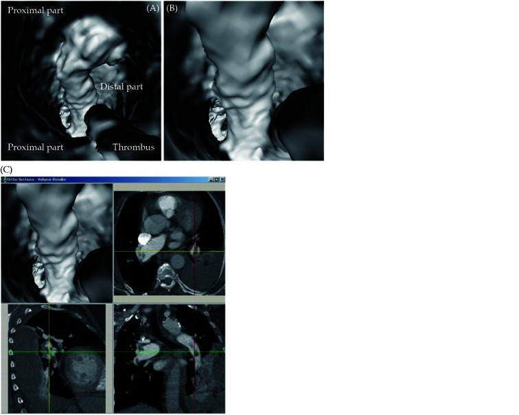
Figure 9.
Virtual intravascular endoscopy views of left lower lobar embolism from proximal to distal segments of lobar artery.
(A): VIE view of proximal segment of left lower lobar pulmonary artery with thrombus; (B): VIE view of distal segment of left lower lobar pulmonary artery with thrombus; (C): accurate position of thrombus is confirmed with using multiplanar views. VIE: virtual intravascular endoscopy. Reprinted with permission under open access from Sun, et al. [68]

