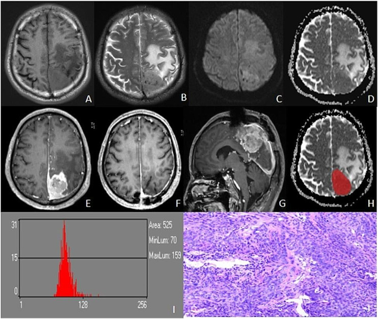
Fig. 3.
Male, 64 years old, left parietal atypical meningioma with recurrence 20 months after surgery. Irregularly shaped iso-T1WI (A) and iso-T2WIsignals in the left parietal, with peritumoural edema (B), slightly hyperintensity in DWI (C), and reduced signals in ADC (D). T1C showed significant inhomogeneous enhancement (E), and there was no recurrence at 3 months after resection (F) and 20 months tumor recurrence (G). Outlined ROI (H), ADC histogram parameters values were as follows: mean, 91.265; variance, 146.88; skewness, -1.3786; kurtosis, 14.874; ADCp1, 70; ADCp10, 81; ADCp50, 91; ADCp90, 103; ADCp99,130 (I). Pathology (HE×100): atypical meningioma, tumor invades brain tissue, multifocal map-like coagulative necrosis, some areas of tightly arranged cells, increased nucleoplasmic proportion of cells, dark staining, pseudo daisy-shaped cluster structure arranged around blood vessels, KI-67% (10–30%) (J)

