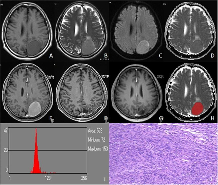
Fig. 4.
Male, 52 years old, left parieto-occipital fibroblastic meningioma with no recurrence till 28 months. The left parieto-occipital region showed a round iso T1WI (A) and slightly longer T2WI signal (B), with signal homogeneity, slightly hyperintensity on DWI (C), and iso-signal on ADC (D). T1C showed significant homogeneous enhancement (E), and there was no recurrence at 3-month (F) and 28-month postoperative review (G). Outlined ROI (H), ADC histogram parameters values were as follows: mean, 94.072; variance, 320.36; skewness, 4.7907; kurtosis, 30.683; ADCp1, 75; ADCp10, 83; ADCp50, 91; ADCp90, 103; ADCp99,184 (I). Pathology (HE×100): fibroblastic meningioma KI-67% was 2% (J)

