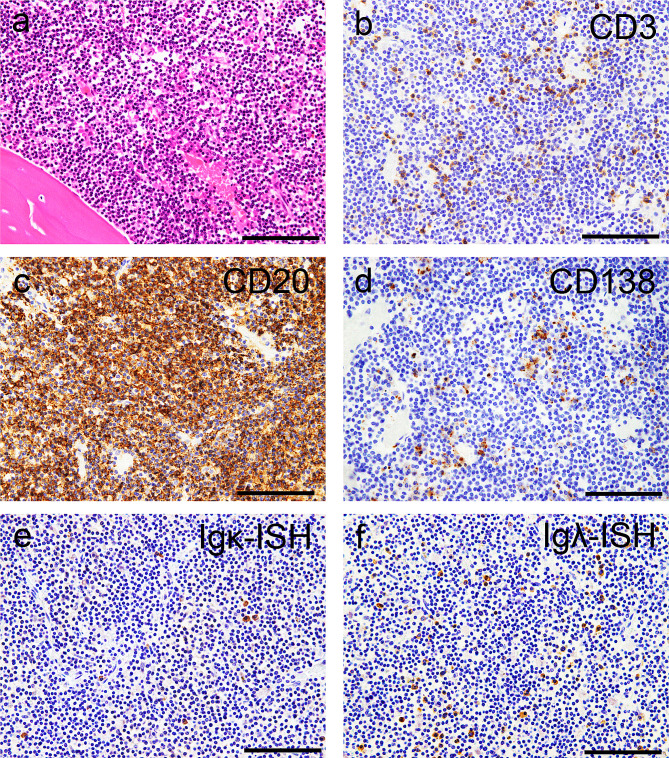
Fig. 1.
Representative microphotographs of the bone marrow. (a) Hematoxylin and eosin staining. (b–d) Immunohistochemistry for CD3 (b), CD20 (c), and CD138 (d). (e, f) In situ hybridization (ISH) for immunoglobulin κ light chain (Igκ) (e) and Igλ (f). (a) Infiltration of lymphocytic cells with small-to-medium-sized nuclei is observed (a). (b, c) The cells are focally positive for CD3 (b) and diffusely positive for CD20 (c). (d) Scattered CD138-immunoreactive cells are also noted. (e, f) Although Igλ-ISH-positive cells are observed more frequently than Igκ-ISH-positive cells, light chain restriction is indefinite in the autopsy materials. Scale bars: 100 μm

