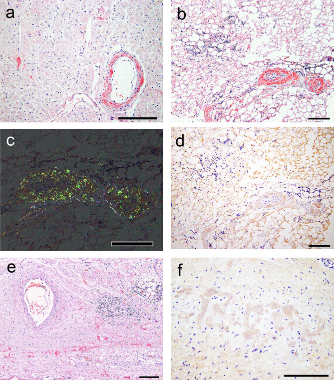
Fig. 3.
Representative microphotographs of the deposits in the heart. (a–c, e) Phenol Congo red (CR) staining under bright field observation (a, b, e, f) and polarized light observation (c). (d, f) Immunohistochemistry for Igλ. (a) Left ventricle. (b–d) Epicardium. (e, f) Sinoatrial node and surrounding atrial tissue. (a) CR-positive deposits are observed in the interstitium and vessels in the left ventricle. (b) More CR-positive deposits are observed in the epicardium than in the myocardium, and lymphoid cell infiltration is also observed. (c) The deposits exhibit apple-green birefringence under polarized light. (d) The amyloid deposits are positive for Igλ. (e, f) Amyloid deposits are observed in the interstitium of the sinoatrial node and show immunoreactivity for Igλ. Scale bars: 200 μm

