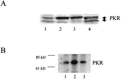
FIG. 1.
(A) Western blot of PKR in infected W138 cells. Cells were mock treated, transfected with dsRNA, or infected with delNS1 or PR8 virus at an MOI of 2. For dsRNA transfection, 50 μg of poly(I)-poly(C) RNA was transfected using 30 μl of DOTAP transfection reagent according to the manufacturer's protocol (Boehringer, Mannheim, Germany). Twenty-four hours postinfection or posttransfection, respectively, cells were lysed, and equivalent amounts of cell extracts were subjected to sodium dodecyl sulfate–polyacrylamide gel electrophoresis. PKR-specific bands were detected by the PKR-specific antibody K-17 (Santa Cruz Biotechnology catalogue no. sc 707; Santa Cruz, Calif.). The upper band corresponds to phosphorylated (active) PKR. The lower band corresponds to unphosphorylated (inactive) PKR (18). The two PKR bands are indicated at the right. Lane 1, mock; lane 2, dsRNA; lane 3, delNS1 virus; lane 4, PR8 virus. (B) Immunoprecipitation of phosphorylated PKR of infected HeLa cells. A total of 106 HeLa cells were mock treated or infected with influenza delNS1 or PR8 virus at an MOI of 0.5. At 5 h postinfection, the cells were washed with a phosphate-free buffer and incubated for 2 h in Dulbecco modified Eagle medium lacking both phosphate and pyruvate (Sigma), containing 500 μCi of [32P]orthophosphate (Amersham). After being labeled, the cells were washed twice with cold phosphate-buffered saline and 10 mM EDTA (without Ca2+ and Mg2+) and lysed for 10 min on ice in lysis buffer. One quarter of the extract was used for immunoprecipitation carried out with 2 μg of PKR antibody B-10 (Santa Cruz Biotechnology catalog no. sc 1215), per ml followed by the addition of 30 μl of protein G-agarose (in a 50/50 ratio) at 4°C. The beads were washed according to the manufacturer's protocol with wash buffer containing PBSTDS (Oncogene, Cambridge, Mass.), heated for 2 min at 95°C and analyzed on a sodium dodecyl sulfate–10% polyacrylamide gel electrophoresis gel. The size of the bands was determined by a size marker (Benchmark, GIBCO-BRL). The bands of phosphorylated PKR were visualized by autoradiography for 7 days and quantified by laser densitometry. Lane 1, mock; lane 2, delNS1 virus; lane 3, PR8 virus. The size marker is indicated at the left. The PKR band is indicated at the right.

