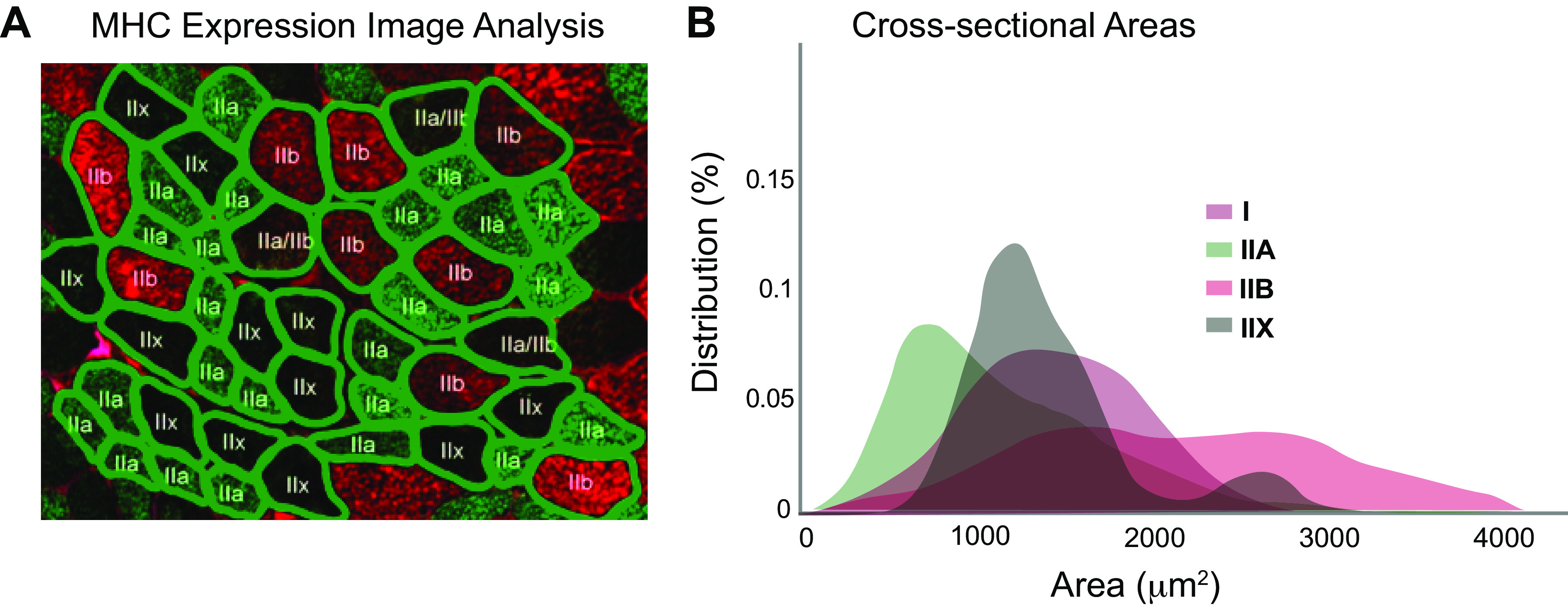
Figure 1.

Illustration of segmentation of ∼50 muscle fibers (A) from one muscle section stained for myosin heavy chain (MHC). An automated method is used to detect fiber boundaries, and fiber types are labeled automatically based on color. This analysis leads to measurement of variation in cross-sectional area across fibers (B), clearly showing significant overlap in size across fiber types. Adapted from Liu et al. (41).
