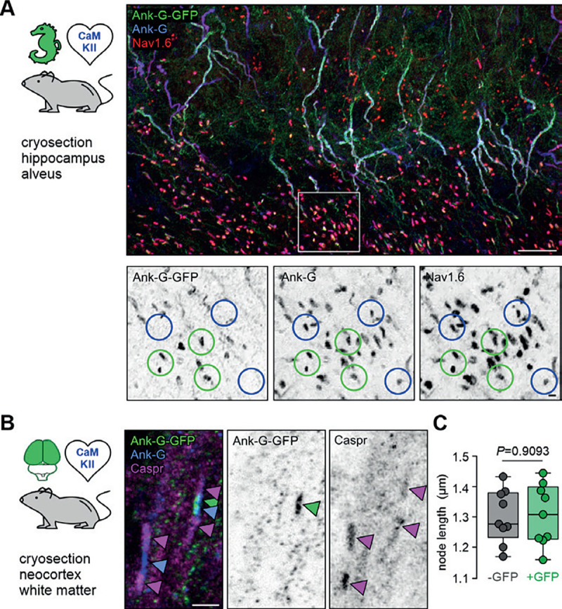
Figure 3. Ank-G-GFP activation and expression in nodes of Ranvier does not alter their morphology.

A Cryosection of hippocampal CA3 alveus from an ank-G-GFP x CaMKIIa-Cre mouse, highlighting ank-G-GFP+ node of Ranvier (noR, green circles) and ank-G-GFP−, but ankyrin-G+ and Nav1.6+ noR (blue circles) in excitatory neurons. Magnification of the region demarked by a white box is shown in inverted black & white panels. All noR express ankyrin-G (middle) and Nav1.6 (left), but only those belonging to CaMKII+ neurons express the ank-G-GFP construct (right). B Cryosection of neocortical white matter from an ank-G-GFP x CaMKIIa-Cre mouse, highlighting ank-G-GFP+ noR (green arrowhead). Ankyrin-G immunoreactivity (blue arrowheads) is seen in both nodes in the image. Caspr is expressed in paranodal regions of both noR (magenta arrowheads). C Quantification of the length of noR using the ankyrin-G signal in control (grey) and ank-G-GFP+ neurons (green) in cortical white matter of ank-G-GFP x CaMKIIa-Cre mice shows no difference between the groups (unpaired t-test, n = 255 nodes in 9 images from 3 animals). Scale bars A = 20 μm, panels in A = 2 μm; B = 2 μm.
