Fig. 5:

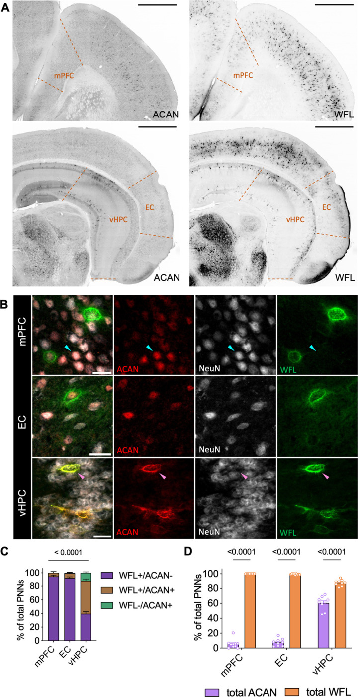
Perineuronal net labeling is highly dissimilar between species and brain region A Representative micrographs of the distribution of either ACAN or WFL labeling across control mouse mPFC, EC and vHPC (N = 10). Imaged at 20X on Evident Scientific VS120 Slide Scanner, scale bar 500μm. B PNNs were manually classified as either single (WFL+/ACAN− or WFL−/ACAN+) or double (WFL+/ACAN+, pink arrow) labeled. As in the human samples, ACAN staining can be found either intracellularly (100% colocalization with NeuN, cyan arrow) or in the distinctive pattern of an extracellular PNN (surrounding NeuN, pink arrow), only the latter was considered as an ACAN+ PNN in this study. Imaged at 40X on Evident Scientific FV1200 Confocal, scale bar 25μm. C PNN labeling pattern varies significantly by brain region (ANOVA: labeling × region F(4,50) = 140.4, p < 0.0001). Unlike the human samples, barely any PNNs were labeled solely by WFL−/ACAN+ in control mouse mPFC and EC. D Examination of total WFL or ACAN labeling within brain regions reveals that WFL labels significantly more PNNs in all brain regions examined (ANOVA: total labeling × region F(2,32) = 330.2, p < 0.0001). WFL: Wisteria Floribunda Lectin, ACAN: anti-aggrecan core protein, mPFC: medial prefrontal cortex, EC: entorhinal cortex, vHPC: ventral hippocampus.
