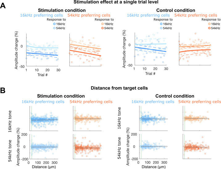
Figure 4. Rebalanced response changes on non-target 16 kHz cells are immediate and widely distributed.
A: Stimulation effect () in 16 kHz (blue) and 54 kHz (orange) preferring cells per each trial for the 5-cell stimulation condition (left) and the no-cell control condition (right). Decreased amplitudes to preferred frequencies were observed from as early as trial 1 with no significant further changes across trials, regardless of frequencies and conditions (sum-of-squares F-test, all p > 0.05). Solid lines indicate fitted curves and dashed lines indicate 95% confidence intervals. B: Stimulation effect () of each non-target 16 kHz (blue) or 54 kHz (orange) preferring cells for either 16 kHz (top row) or 54 kHz (bottom row) presentation in relation to the minimum distance to any target cells for the stimulation condition (left) and the control condition (right). For the control condition, we considered top 5 most tone-responsive cells from the baseline session as “target” cells, as there was no stimulation. Non-target cells are widely distributed within the FOV (550 μm2), regardless of cell groups, frequencies, and conditions. Gray lines indicate fitted curves, excluding cells that are closer than 15 μm (vertical green lines; cells < 15 μm marked in lighter shades), and dashed lines indicate 95% confidence intervals.

