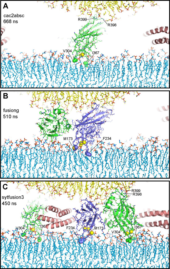
Figure 2.
Syt1 C2 domain Ca2+-binding loops do not insert deeply into lipid bilayers and cause limited bilayer perturbation. The diagrams show examples of C2 domains with their Ca2+-binding loops interacting with the flat bilayer from frames taken at 668 ns of the cac2absc simulation (A), 510 ns of the fusiong simulation (B) and 450 ns of the sytfusion3 simulation (C). Lipids are shown as stick models. Ca2+ ions are shown as yellow spheres. SNARE complexes are represented by ribbon diagrams and Syt1 C2 domains by ribbon diagrams and stick models with nitrogen atoms in dark blue, oxygen in red, sulfur in yellow orange and carbon colored in slate blue (C2A) and green (C2B). Other color coding is as in Fig. 1. The hydrophobic residues at the tips of the Ca2+-binding loops that insert into the flat bilayer are shown as spheres and labeled. R398 and R399 at the opposite end of the C2B domain are labeled in (A, C) to illustrate how the C2B domain can bridge two membranes as predicted (42).

