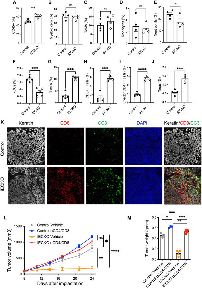
Figure 2. Abrogation of endothelial Notch signaling selectively promotes the infiltration of T cells in PDAC models.
A-J. Flow cytometric quantification of indicated leukocyte populations in subcutaneous KP1 tumors of control and RbpjiECKO mice. Representative of >5 independent experiments.
K. Immunofluorescence imaging of CD8 (red), cleaved caspase 3 (green), and keratin (white) in KP1 tumors from control and RbpjiECKO mice, visualized by the 10X objective.
L-M. Volumetric (L) and weight (M) measurements of subcutaneous KP1 tumors in control and RbpjiECKO mice treated with CD4/CD8 depleting antibodies (n=3–6/group). Representative of >3 independent experiments.
* p<0.05, ** p<0.01, *** p<0.001, **** p<0.0001 by student’s t-tests. Ns denotes not significant.

