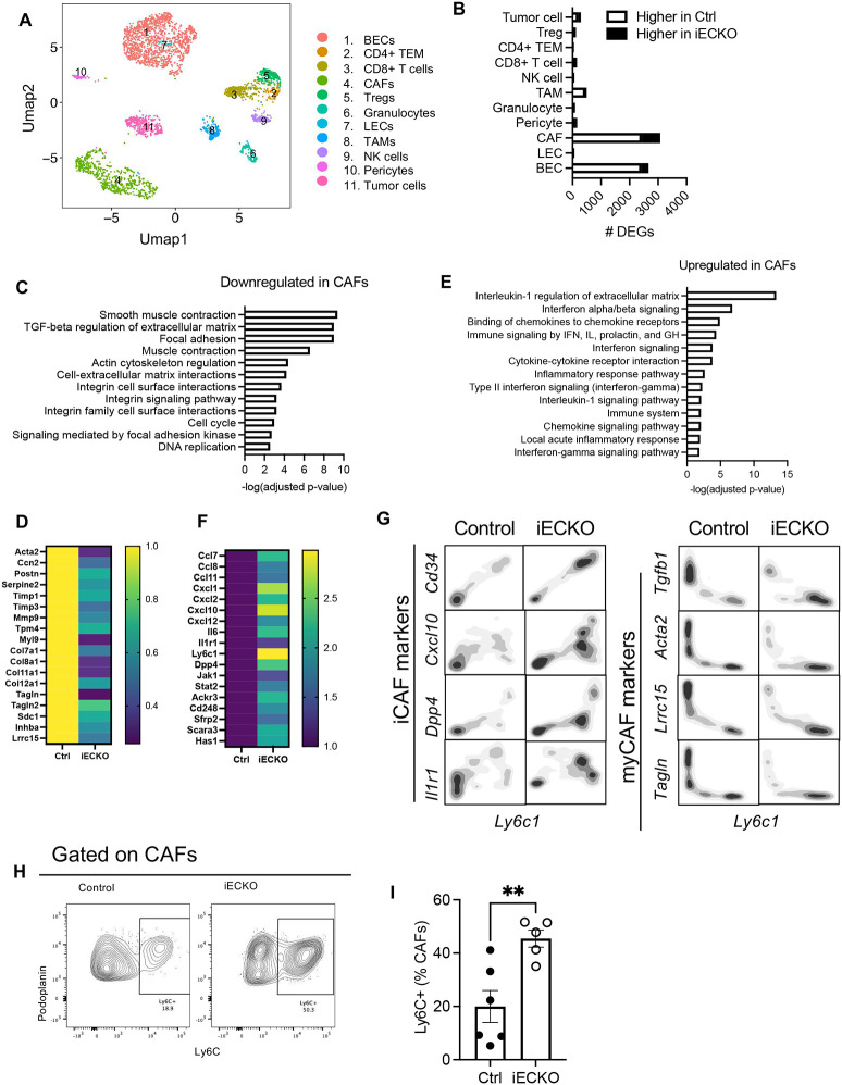
Figure 3. Abrogation of endothelial Notch signaling inhibits myofibroblastic CAFs and promotes pro-inflammatory CAF signature.
A. Umap plots of tumor and stromal cells by scRNAseq analyses of PDAC in control and RbpjiECKO mice.
B. Number of differentially expressed genes (DEGs) in indicated cell types in response to RbpjiECKO, (p-value <0.01).
C-D. BioPlanet pathway analyses (C) and heatmap (D) of DEGs in CAFs downregulated by RbpjiECKO.
E-F. BioPlanet pathway analyses (E) and heatmap (F) of DEGs in CAFs upregulated by RbpjiECKO.
G. Contour plot of scRNAseq of CAFs as marked by indicated molecules.
H-I. Representative plots (H) and mean fluorescence intensity (MFI) quantification (I) of Ly6C expression in CAFs from KP1 tumors in control and RbpjiECKO mice. Representative of >5 independent experiments.* p<0.05 by student’s t-tests.

