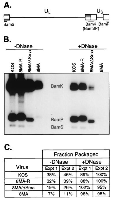
FIG. 2.
DNA encapsidation assay. (A) Schematic diagram of the HSV-1 genome. The unique long (UL) and unique short (US) regions are flanked by the long and short inverted repeats (hatched and open boxes, respectively). Digestion by BamHI within these repeats results in the indicated BamHI restriction fragments. (B) Southern blot analysis of HSV-1 DNA. Infected Vero cell lysates were harvested, either treated or left untreated with DNase I, and subjected to SDS and proteinase K treatment. DNA was subsequently extracted for BamHI digestion. The locations of the BamHI K, S, and P restriction fragments are indicated. (C) Quantitation of the fraction of packaged DNA. The intensities of the BamHI K, S, and P fragments from panel B (experiment [Expt] 1) were quantitated by phosphorimager analyses, and the fraction of packaged viral DNA was determined. Quantitation of an independent experiment not illustrated in panel B is also shown (experiment 2).

