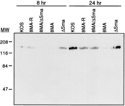
FIG. 3.
Western blot analysis of HSV-1-infected Vero cells. Vero cells were infected with the indicated viruses at an MOI of 5 and harvested into SDS-PAGE lysis buffer at 8 or 24 h postinfection. The major capsid protein, VP5, was visualized by using a 1:20,000 dilution of NC-1. The location of molecular mass markers are indicated in kilodaltons.

