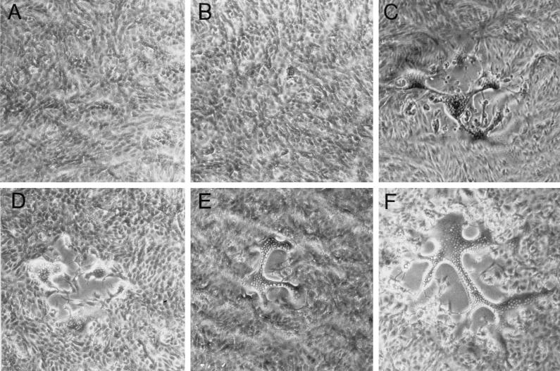
FIG. 9.
Plaque morphology of HSV-1 recombinants. Vero cells were infected with 8MA (A), 8MA/ΔSma (B), 8MA-V (C), 8MA/UL53syn (D), 8MA/ΔSma/UL53syn (E), and KOS/UL53syn (F) under limiting dilutions in order to visualize individual plaques. Monolayers were photographed at ×40 magnification 2 days postinfection. 8MA and 8MA/ΔSma fail to plaque on Vero cells, while the remaining recombinant viruses form syncytial plaques where individual nuclei can be visualized within a syncytium.

