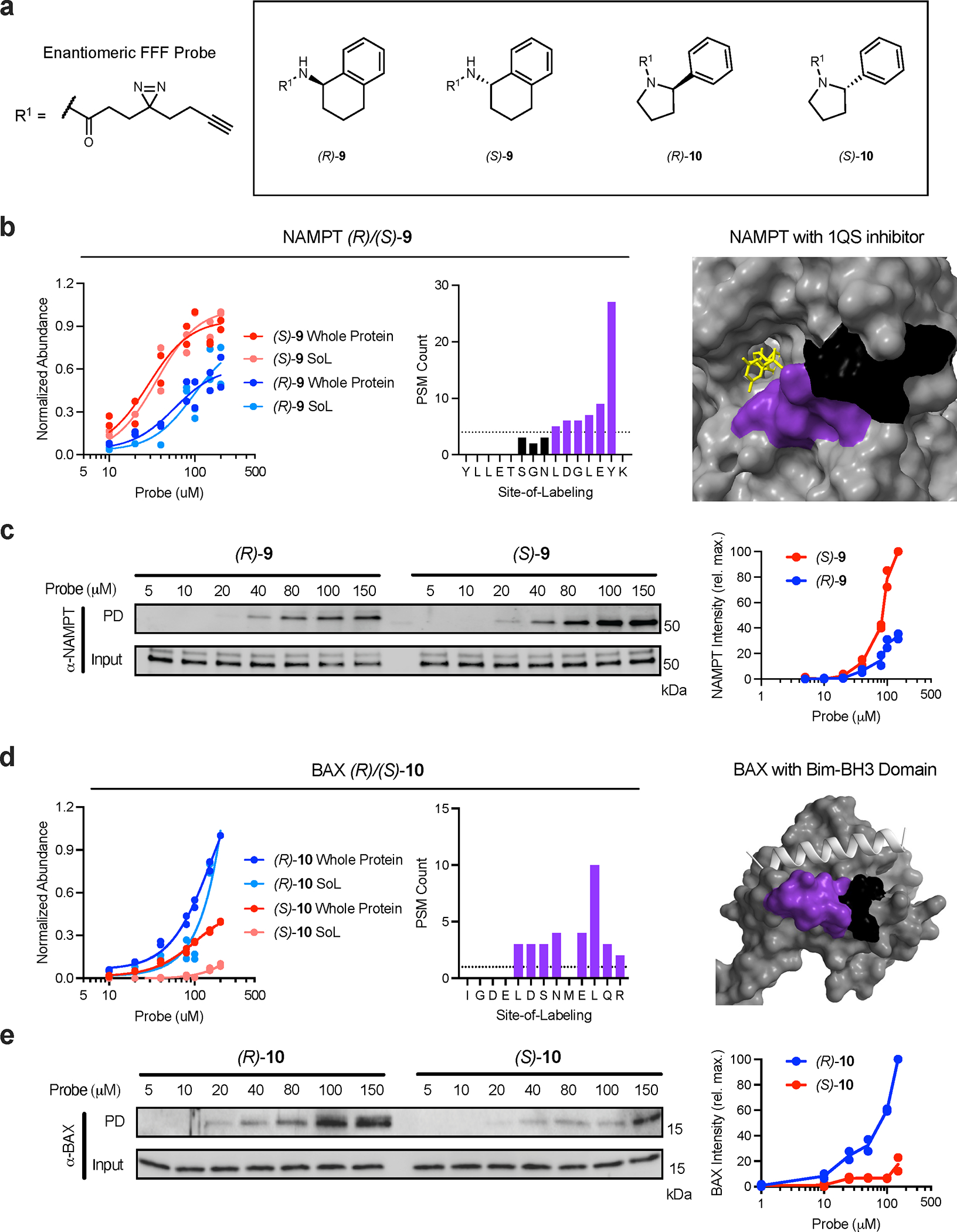
Figure 5.

Proteome-wide, site-specific, concentration-dependent, probe-protein interaction profiles. For proteomic experiments, cells were treated with seven different concentrations (10, 20, 40, 80, 100, 150, 200 μM) of FFF probes before being UV crosslinked and processed as indicated in Fig 4a. For all structures, the primary peptide residues labeled by probes are colored purple and the remainder of each peptide is colored black. Co-resolved ligands are colored yellow and other proteins are colored silver. (a) Structures of enantiomeric FFF probes used in the TMT dose experiments. (b) NAMPT concentration plot and probe label site overlapping with known inhibitor (PDB: 4KFO). (c) Immunoblot analysis and quantification of NAMPT stereoselective probe binding. (d) BAX concentration plot and probe label site proximal to the active site (PDB: 4ZIE). (e) Immunoblot analysis and quantification of BAX stereoselective probe binding. Each immunoblot displayed is representative of two independent experiments. (PD = pulldown)
