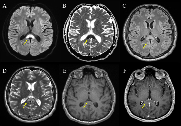
Fig. 2.
Typical structural MRI findings in cytotoxic lesions of the corpus callosum (CLOCC). Pictorial magnetic resonance imaging (MRI) examples of a 38-year-old patient who presents with a typical cytotoxic lesion of the corpus callosum (CLOCC) after head trauma. A DWI B0, oval hyperintensity throughout the splenium and into the adjacent hemispheres (“boomerang sign”). B ADC hypointensity due to restricted diffusion. C T2w-FLAIR, high signal. D T2 slightly hyperintense. E T1 native, pre-gadolinium uptake, shows a slight hypointensity in the region. F T1 post-gadolinium uptake, no enhancement. Abbreviations. DWI, diffusion-weighted imaging; FLAIR, fluid-attenuated inversion recovery

