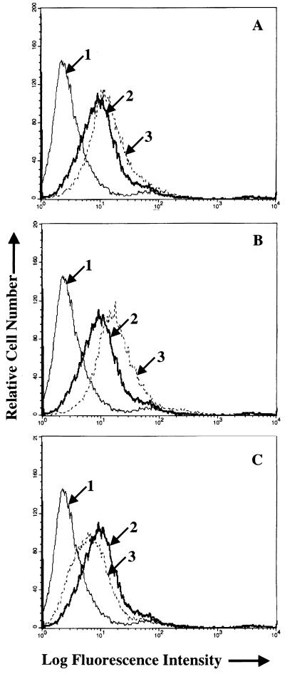
FIG. 6.
Flow-cytometric analysis of the effects of MAbs CL40 (A), CL59 (B), and E1D1 (C) on binding of wild-type Akata virus to AGS cells. In each panel arrow 1 indicates the histogram of cells incubated with MAb to 72A1 to gp350 and fluorescein-conjugated sheep anti-mouse immunoglobulin alone; arrow 2 indicates the virus bound after preincubation with phosphate-buffered saline and visualized with MAb 72A1 and fluorescein-conjugated sheep anti-mouse immunoglobulin; arrow 3 indicates the virus bound after preincubation with MAb C140, CL59, or E1D1 and visualized with MAb to 72A1 and fluorescein-conjugated sheep anti-mouse immunoglobulin.

