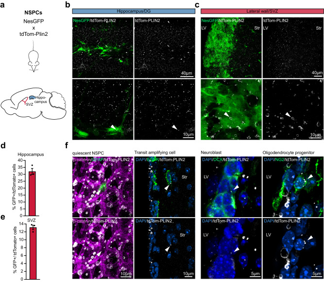
Fig. 5. LDs are present in NSPCs and their progeny in the postnatal and adult mouse brain.
a Scheme of the crossing of tdTom-Plin2 mice to NesGFP reporter mice, labelling NSPCs in the SVZ and DG. b, c Endogenous tdTom-PLIN2 signal in the DG and SVZ showing LDs in NSPCs. Representative images of non-stained sections show NesGFP positive NSPCs and tdTom-PLIN2 positive LDs. (Maximum intensity projections,10 μm stacks). d FACS analysis of cells isolated from the hippocampus of tdTom-Plin2 mice crossed to NesGFP mice show that around 30% of the NesGFP positive NSPCs have LDs. (n = 3 mice, mean +/- SEM). e FACS analysis of cells isolated from the SVZ of tdTom-Plin2 mice crossed to NesGFP mice show that around 13% of the NesGFP positive NSPCs have LDs. (n = 3 mice, mean +/- SEM). f Immunohistochemical staining of cells in the SVZ show tdTom-PLIN2 positive LDs in GFAP positive quiescent NSPCs (whole mount), EGFR positive transit amplifying cells, DCX positive neuroblasts and NG2 positive oligodendrocyte progenitors (coronal sections). Representative maximum intensity projections of the observed cell types. (n = 3 mice, similar observations in at least 10 cells per cell type and mouse).

