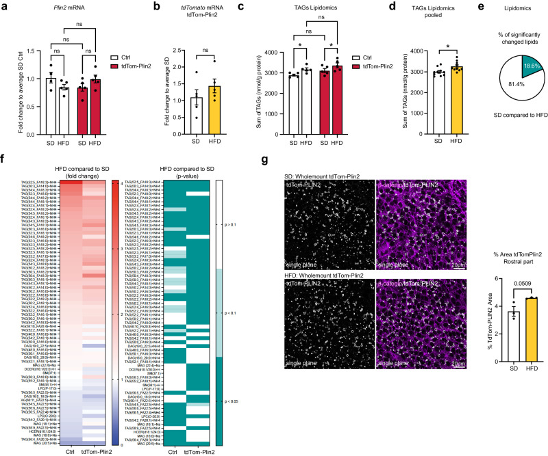
Fig. 6. Short-term high fat diet slightly increases the total TAG levels in the brain and moderately increases LDs in the wall of the lateral ventricles.
a Analysis of mRNA expression by RT-qPCR shows no significant difference in Plin2 due to the HFD in either Ctrl mice or tdTom-Plin2 mice. (n = 4 Ctrl SD, 5 Ctrl HFD, 5 tdTom-Plin2 SD, 5 tdTom-Plin2 HFD mice), fold change +/- SEM). b tdTomato was also not altered on the gene expression level in SD or HFD tdTom-Plin2 mice (n = 5 mice per group, fold change +/- SEM). c Lipidomic analysis revealed an increase in brain TAGs upon HFD compared to SD, (n = 5 mice per group, mean +/- SEM). d Pooling of genotypes shows that TAGs are significantly increased in HFD fed mice compared to SD mice, however the increase is overall very moderate. (n = 10 mice per group, mean +/- SEM). e Out of 366 analysed lipids in Ctrl and tdTom-Plin2 brains by lipidomics, 18.6% were significantly different when comparing SD to HFD. f The majority of the significantly changed brain lipids were TAGs. When looking at their fold changes, both Ctrl and tdTom-Plin2 brains showed a coherent pattern in terms of increased and decreased lipid species as a reaction to the HFD (left heatmap shows fold change to SD, right heatmap shows the corresponding p-values). g Quantification of the area covered by tdTom-Plin2 signal in whole mount preparations of the lateral ventricle wall shows a slight increase in LDs with HFD in the rostral part. Representative single planes, β-catenin outlines the cell membranes of ependymal cells. (n = 3 mice per group, mean +/- SEM). Asterisks indicate the following p-values: *< 0.05. ns = non-significant.

