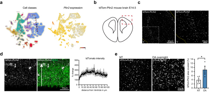
Fig. 7. LDs are present in the developing embryonic brain and react dynamically to exogenous lipids.
a Querying for Plin2 expression in scRNA sequencing data from embryonic brain development (http://mousebrain.org) shows that Plin2 is abundant in many clusters, one of them being radial glia (RG) cells. b Illustration of a coronal section from an embryonic brain at E14.5, red square marking the dorsal pallium, where LDs were imaged. c tdTom-PLIN2 positive LDs are abundant in the developing cortex at E14.5 in tdTom-Plin2 embryos. (Maximum intensity projection, 25 μm stack, at low and high magnification). d Quantification of tdTomato intensity in relation to distance from the ventricle shows that LDs are homogenously distributed throughout the Sox2-positive layer. Arrow indicates the direction of measurement (n = 3 embryos, mean +/- SEM). e Overnight incubation of OA significantly increases the LD area covered compared to non-treated (NT) sections (n = 4 embryos, mean +/- SEM). *< 0.05.

