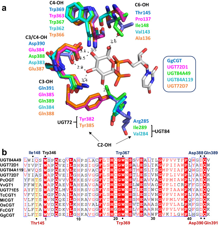
Fig. 6. Active site close-ups and partial multiple sequence alignment showing the conserved donor binding residues in UGTs.
a The donor complex of GgCGT74 (blue carbons; PDB: 6L5P) with UDP-Glc (gray carbons) overlaid with the AlphaFold-predicted structures of UGT72D1 (pink), UGT84A49 (light green), UGT84A119 (dark cyan) and UGT72D7 (orange). The conserved tetrad Gln-Asp/Glu-Trp-Thr for the binding of glucose C2-OH, C3-OH, C4-OH and C6-OH is seen in GgCGT. b Sequence comparison of flavonoid O- and C-glycosyltransferases indicating the UDP-Glc binding interactions in GgCGT (red, below the alignment) and UGT84A49 (blue, above the alignment). The conserved Thr for stabilization of glucose C6-OH is highlighted in yellow, for UGT84A49 and UGT84A119 the corresponding residue is Ile/Val in the preceding position in the sequence. The black numbering corresponds to the positions in the PSPG box, as show in Fig. 2a.

