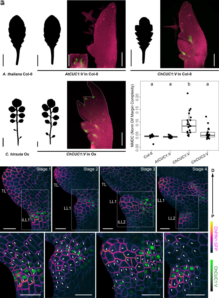
Fig. 1.
ChCUC1 is sufficient to increase A. thaliana leaf complexity upon interspecific gene transfer and its expression associates with PIN1 polarity reversals. (A and B) Silhouettes of rosette leaf 8 from wild-type A. thaliana (Columbia-0, Col-0) (A) and C. hirsuta (Oxford; Ox) (B). (C and D) Silhouettes (C) and transgene expression (D) in A. thaliana rosette leaf 8 carrying AtCUC1:V (AtCUC1p::AtCUC1g:Venus). The Inset in (D) shows the vegetative shoot apex. (E and F) Silhouettes (E) and transgene expression (F) in C. hirsuta rosette leaf 8 carrying ChCUC1:V (ChCUC1p::ChCUC1g:Venus). (G and H) Silhouettes (G) and transgene expression (H) in A. thaliana rosette leaf 8 carrying ChCUC1:V (ChCUC1p::ChCUC1g:V). (D, F, and H) Maximum intensity projections of confocal stacks. Green: Venus signal; magenta: cell walls visualized with propidium iodide (PI). (I) Leaf complexity of leaf 8 from the indicated genotypes by measurement of Normalized Difference Margin Complexity [(perimeter contour-perimeter convex hull)/(perimeter contour + perimeter convex hull)]. Letters a and b indicate significant differences by the Kruskal–Wallis test with Dunn’s post hoc test (α = 0.05). ChCUC2:V refers to ChCUC2p::ChCUC2g:V (34). (C–I) Replication: n (phenotypic analysis) ≥ 20 transgenic T1 lines, n (confocal microscopy) = 3 lines. (J–M’) C. hirsuta leaf 5 at different developmental stages showing epidermal expression of ChCUC1:V (ChCUC1p::ChCUC1g:V) and ChPIN1:GFP (ChPIN1p::ChPIN1g:eGFP) projected onto a MorphographX mesh (SI Appendix, Materials and Methods). The white arrows on the leaf margin cells indicate the direction of ChPIN1:GFP polarity. The black arrowheads in (L’) indicate leaf margin cells with ChCUC1:V expression and apical ChPIN1:GFP polarity. The dotted Insets indicate the areas magnified in (J’–M’). D, distal; P, proximal; TL, terminal leaflet; LL, lateral leaflet; iLL, initiating lateral leaflet. n = 3 leaves per stage. The leaf silhouettes were obtained 21 d after sowing. [Scale bars: 1 cm (A–C, E, and G); 100 µm (D, F, and H); 20 µm (J–M’).]

