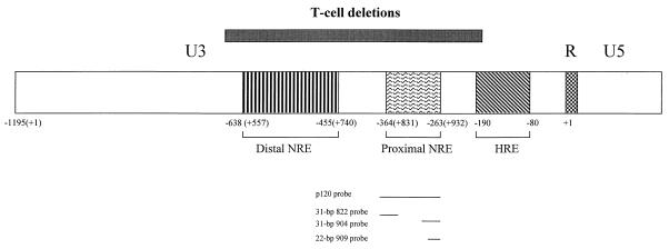
FIG. 1.
Diagram of the MMTV LTR. The LTR is divided into U3, R, and U5 regions, and transcription is initiated from the standard MMTV promoter at the first base of the R region. The promoter-proximal and promoter-distal NREs and the HRE are shown by boxes with different types of hatch marks within the U3 region of the LTR. Numbering is shown from the first base of the LTR (+1). The region encompassing the largest of the U3 deletions found in thymotropic MMTVs relative to mammotropic MMTV strains is shown by a box over the LTR. The positions of probes used in this study are given below the LTR. The number of bases in each probe also is indicated.

