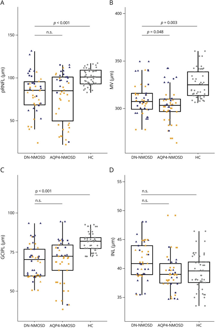
Figure 1. Retinal Layer Thicknesses in DN-NMOSD, AQP4-NMOSD, and HCs.

Box plots with dotted overlay for single-eye–based values for eyes with a history of ON (orange) and without a history of ON (blue) as well as for HC eyes (gray). Dots are shaped depending on the history of contralateral ON (history of contralateral ON: square, no history of contralateral ON: triangle, HC: circle). AQP4-NMOSD = people with aquaporin-4 antibody seropositive neuromyelitis optica spectrum disorder; DN-NMOSD = people with double-antibody seronegative neuromyelitis optica spectrum disorder; GCIPL = combined ganglion cell and inner plexiform layer; HC = healthy control; INL = inner nuclear layer; MV = macular volume; n.s. = not significant; ON = optic neuritis; pRNFL = peripapillary retinal nerve fiber layer.
