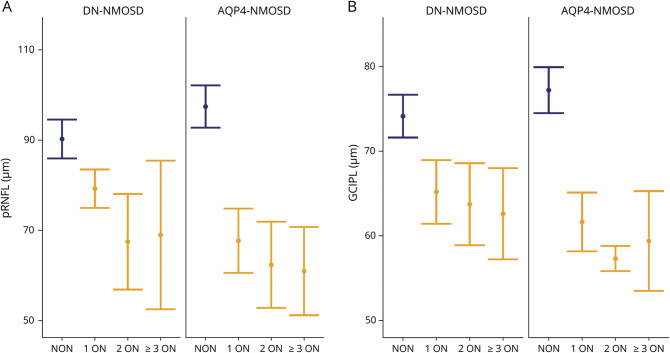
Figure 2. Retinal Neurodegeneration After ON in DN-NMOSD and AQP4-NMOSD.
Mean layer thickness (dot) with standard error of the mean (whiskers) for eyes without ON (NON, blue) as well as for eyes after 1 ON, 2 ON, and ≥3 ON (orange) of people with DN-NMOSD (left) and AQP4-NMOSD (right). Owing to the low sample size, none of the within-group comparisons (NON vs 1 ON, 1 ON vs 2 ON, 2 ON vs ≥3 ON) was statistically significant. AQP4-NMOSD = people with aquaporin-4 antibody seropositive neuromyelitis optica spectrum disorder; DN-NMOSD = people with double-antibody seronegative neuromyelitis optica spectrum disorder; GCIPL = combined ganglion cell and inner plexiform layer; ON = optic neuritis; pRNFL = peripapillary retinal nerve fiber layer.

