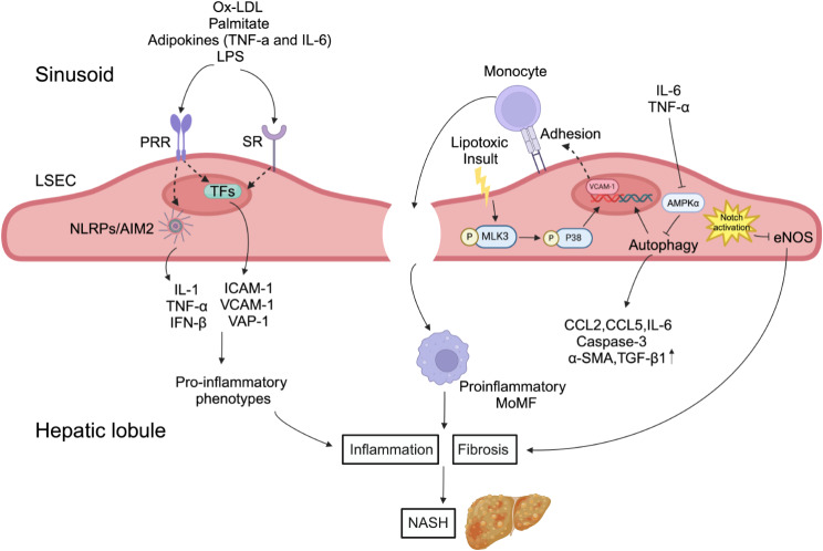
Fig. 4.
Ox-LDL, palmitate, adipokines (TNF-a and IL-6) and LPS lead to inflammatory phenotypes in LSECs, which induce LSECs to produce pro-inflammatory mediators in MASH, including TNF-α, IL-6, IL-1 and CCL2. Lipotoxic stress can enhance the VCAM-1 expression of LSECs through MLK3/P38 signal transduction, and stimulate monocytes to gather in the liver through fenestrae and activate into a pro-inflammatory state. In addition, IL-6 and TNF-α in local microenvironment down-regulate autophagy and increase the expression of CCL2, CCL5, IL-6, Caspase-3, α-SMA and TGF-β1 by inhibiting AMPKα, and activation of Notch signal in damaged LSECs inhibits eNOS transcription, which aggravates liver inflammation and fibrosis, and then promotes the progression of MASH. MOMF: monocyte-derived macrophages

