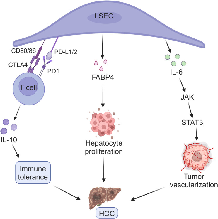
Fig. 6.
LSECs related pathological mechanism of HCC. The PD-L1/2 and CD80/86 expressed on LSECs constitute the ligands of PD1 and CTLA4 in T cells, respectively. In the interaction, the activation of T cells is inhibited and a state of tolerance differentiation promoted by locally produced IL-10 is obtained. In the microenvironment, LSECs exposed to high concentration of glucose, insulin or VEGF-A can induce hepatocyte proliferation and promote HCC growth by releasing FABP4. In addition, IL-6 and IL-6 receptor secreted by peritumoral endothelial cells and macrophages lead to highly vascularized tumors, which simultaneously aggravate the development of tumor occurrence

