
Figure 1. Physiological inspiration of bioprinting salivary gland ductal and epithelial features.
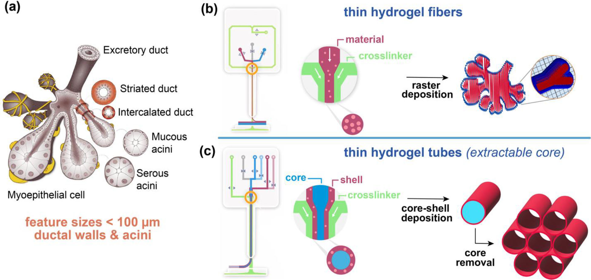
(a) Scheme of fully developed salivary gland (adopted from Ref [37]). (b) Scheme of DUO Printhead of RX1 bioprinter and the raster deposition method using solid fibers to print biomimetic structures. (c) Scheme of CENTRA Printhead of RX1 bioprinter and a core-shell deposition method to produce hollow tubes with thin walls.