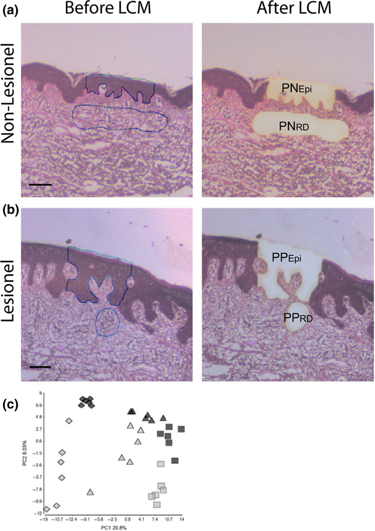
Figure 1.
Combined use of laser capture microdissection (LCM) and small RNA sequencing identifies specific miRNA signatures in psoriatic plaque epidermis and dermis. LCM was performed on both normal psoriatic skin (a) and psoriatic plaque (b) samples. Cells were collected from psoriatic epidermis (PNEpi and PPEpi) and reticular dermis (PNRD) which included inflammatory cell infiltrates in lesional samples (PPRD). (a, b) Images are representative for 6 biopsies. Line equals 100 μm. A PCA plot based on the normalized read frequencies of all samples clearly showed a separation of the samples according to (c) the sample type (rhombus = PNEpi+PPEpi, triangle = whole-tissue extract, square = PNRD+PPRD) and indication (grey = psoriatic plaque, black = normal psoriatic skin).

