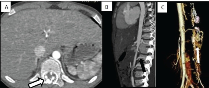
Figure 1.
A–C Axial and sagittal CT angiography and volume rendered images showing aneurysmal enhancing dilated tortuous vascular channels (black outlined arrow), multiple arterial feeders as the thoracic and lumbar radicular arteries supplying the fistulae (solid black arrow). 3D volume rendering technique image in Figure 1C depicts the communication between the feeders and the extensive peri medullary plexus of veins (thin arrow).

