Figure 2.
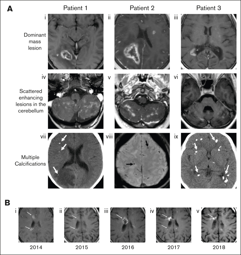
Characteristic radiographic findings of FANS and their evolution over time. (A) Representative MRI findings shown for 3 patients with FA in our cohort. (Ai-iii) Axial postcontrast T1-weighted MRIs show focal ring-enhancing mass lesions. (Aiv-vi) Contrast-enhanced images through the cerebellum show multiple enhancing lesions. (Avii,ix) Noncontrast computed tomography and (Aviii) susceptibility weighted MR images demonstrate multiple calcifications in each case (arrows). (B) Axial T1-weighted MR images from patient 3 over a 5-year span, demonstrating the waxing and waning of lesions. (Bi) In 2014, enhancing lesions are seen on the anterior and posterior margin of the right lateral ventricle (arrows), both of which markedly regress a year later (Bii, arrows). (Biii) In 2016, the anterior lesion recrudesces (arrow), then decreases again over the next 2 years, with some waxing and waning of the posterior lesion (Biv-v, arrows).

